Examination and Imaging
Periapical, Panoramic, Tomography
Success in dental health treatments begins with correct diagnosis. Therefore, the first step is a comprehensive examination and an evaluation process supported by modern imaging techniques. At Dr. Dt. Gizem Ömeroğlu's practice, patients are evaluated in detail with both clinical examination and digital radiological methods. This way, not only current problems but also potential future risks can be detected in advance. The advanced imaging techniques used ensure that the treatment plan is personalized and based on scientific foundations.
Periapical X-ray: Sharp Imaging for Spot Diagnosis
Periapical x-ray allows detailed examination of the root and surrounding tissues of a single tooth or a few teeth. It is particularly preferred in cases of root canal infections, cyst formations, depth of caries and when root canal treatment is needed. This imaging method is of great importance for early diagnosis. Many problems that cannot be noticed from the outside can be detected with periapical x-ray. This method, which contains minimal radiation, contributes to the safe and correct progression of the treatment process.
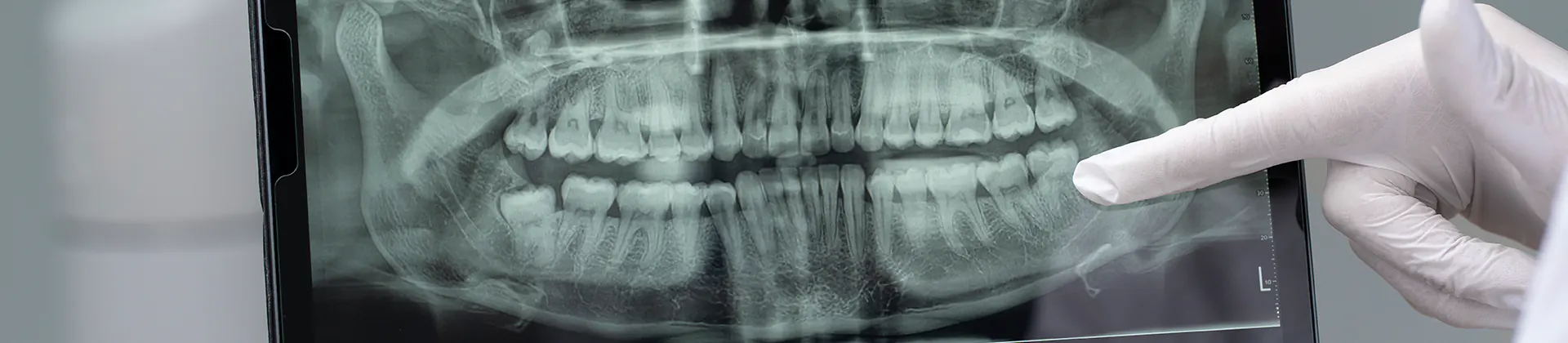
Panoramic X-ray: General Map of Oral and Jaw Structure
Panoramic x-ray presents all teeth in the lower and upper jaw, jaw bones, joint structures and sinus regions in a single image. Thanks to this broad perspective, impacted teeth, jaw bone structure, tooth alignment and potential anomalies can be easily detected. It is frequently preferred for orthodontic treatment planning, monitoring of impacted wisdom teeth and general oral health check-ups. Additionally, thanks to the quick and comprehensive image provided by panoramic x-ray, a more accurate personalized treatment plan can be created for the patient.
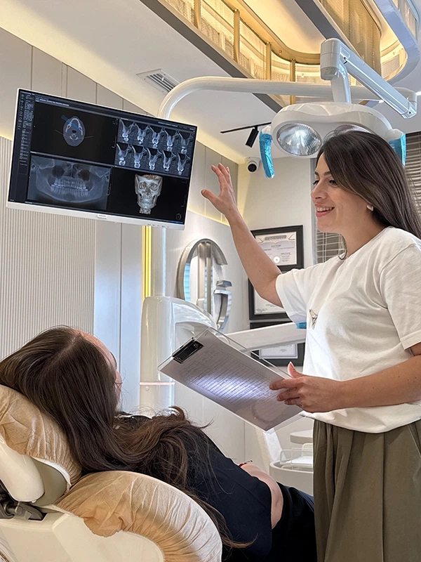
Dental Tomography (CBCT): Three-dimensional and In-depth Examination
Cone Beam Computed Tomography (CBCT) is a revolutionary technology in dentistry. Compared to traditional two-dimensional x-rays, dental tomography provides three-dimensional and much more detailed images. It is an indispensable method in cases requiring information about implant planning, measurement of jaw bone density, evaluation of sinus cavities and complex anatomical structures. Thanks to CBCT, not only the bone structure but also surrounding tissues can be examined in detail. This both increases the success rate of surgical procedures and minimizes complication risks.